Mapping the Mind: Indian Researchers Unveil World’s Most Detailed 3D Brainstem Atlas
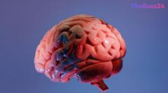
Researchers at the Sudha Gopalakrishnan Brain Centre (SGBC) at the Indian Institute of Technology, Madras (IIT-M), have achieved a significant milestone in neuroscience by developing the world’s most detailed three-dimensional atlas of the human brainstem. Known as Anchor (Atlas of Neurochemical Characterisation of the Human Brainstem with 3D Reconstruction), the project provides a digital map that allows scientists to navigate seamlessly from whole-brain MRI scans down to the level of individual nerve cells. This breakthrough addresses a long-standing challenge in the field, where researchers have historically struggled to bridge the gap between macroscopic imaging and microscopic cellular pathology.
The brainstem, while physically small, is critical for sustaining life, as it regulates essential functions such as breathing, heart rate, and sleep. Despite its importance, its complex, densely packed architecture has made it difficult to map in detail. The Anchor atlas overcomes this by integrating over 500 tissue sections from fetal, childhood, and adult brains, utilizing high-resolution microscopy to identify more than 200 clusters of brain cells and nerve pathways. By applying eight chemical markers, the team has created a clear, navigable reference tool that is now freely available to the global scientific community.
This initiative represents a shift in modern neuroscience, where progress is increasingly driven by the convergence of engineering, computation, and biology. By enabling researchers to compare healthy brainstem structures with those affected by neurological conditions—such as Alzheimer’s disease, Parkinson’s, and stroke—the atlas offers a new lens through which to study disease progression. The SGBC team plans to expand this work by imaging over 100 additional human brains, aiming to build a comprehensive library that could eventually reveal how various disorders reshape the brain at a cellular level.
Key Takeaways
- The Anchor atlas provides a 3D, cellular-resolution map of the human brainstem, bridging the gap between whole-brain MRI scans and microscopic tissue analysis.
- The project utilizes high-resolution microscopy to identify over 200 cell clusters and nerve pathways, offering a vital tool for studying neurological disorders.
- The atlas is freely available online, serving as a global resource for neuroscientists and neurosurgeons to improve diagnostic understanding and surgical precision.
Editor’s Analysis & Impact
The development of the Anchor atlas marks a pivotal moment in neuro-mapping, signaling a transition toward ‘big data’ approaches in biological research. By democratizing access to high-resolution cellular data, this project lowers the barrier for researchers worldwide to conduct comparative studies on neurodegenerative diseases. The industry impact is profound: it moves the field away from isolated, small-sample studies toward a standardized, digital reference framework. Future implications include more precise neurosurgical navigation and a deeper understanding of how systemic conditions, such as post-viral neurological damage, manifest at the cellular level. As the SGBC scales its library to include hundreds of brains, this atlas will likely become a cornerstone for pharmaceutical and clinical research, potentially accelerating the development of targeted therapies for conditions that have historically been difficult to map and treat.
Frequently Asked Questions
Q: What is the primary function of the Anchor atlas?
A: The Anchor atlas serves as a high-resolution, 3D digital map that allows scientists to zoom from whole-brain MRI views down to individual nerve cells within the brainstem, helping to visualize complex structures that were previously difficult to study.
Q: Is the Anchor atlas intended to be used as a diagnostic tool?
A: No, the atlas is not a diagnostic tool. Instead, it is a reference resource designed to help researchers and clinicians better understand the cellular architecture of the brain and how it changes during various neurological diseases.
AI Disclosure: This article is based on verified data and official reports. Our Team and AI have cross-referenced every financial detail with primary sources to ensure total accuracy.
More from this Category